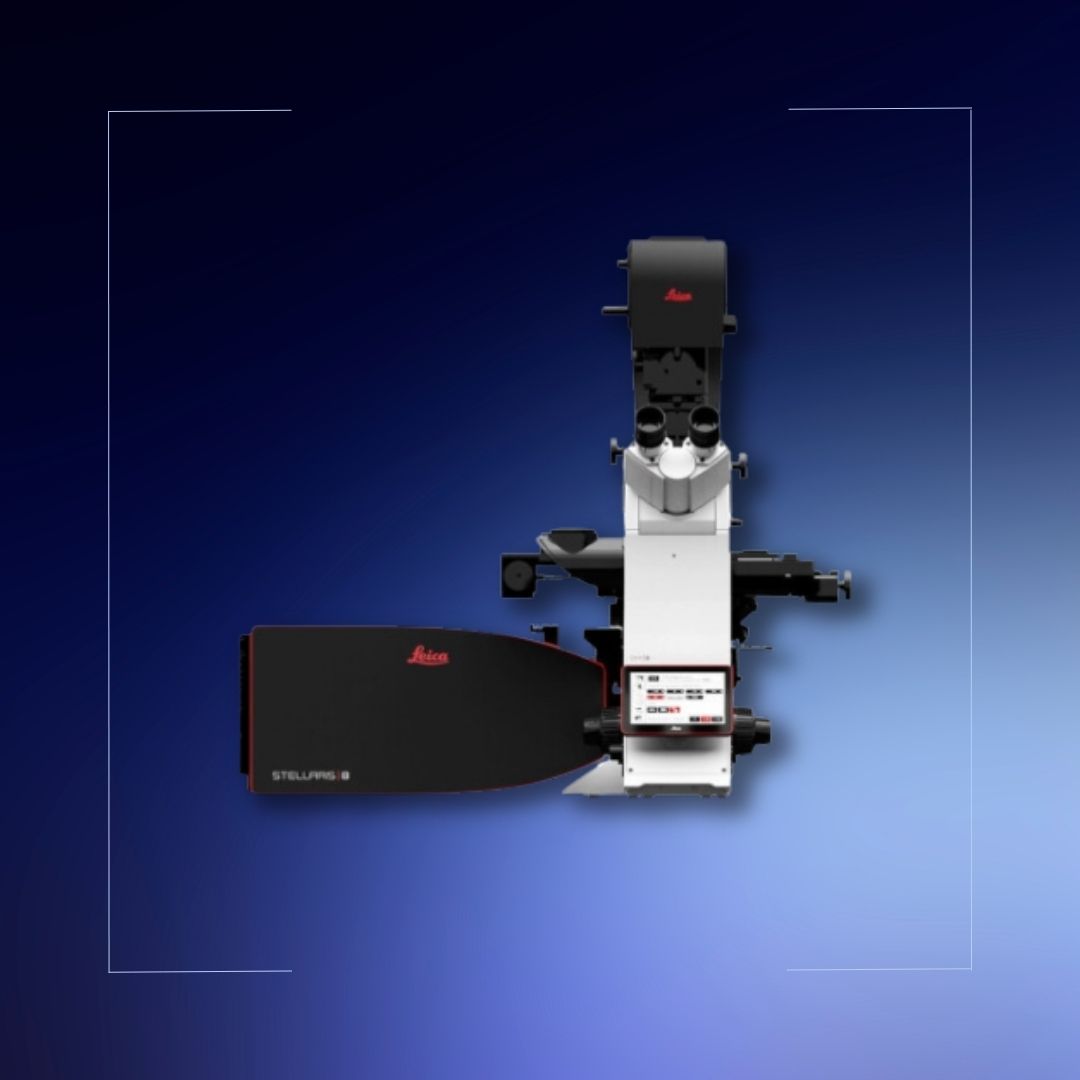
The imaging system combines confocal and digital light-sheet (DLS) microscopy in a single location, enabling images of unparalleled quality to be taken according to experimental needs without hardware modification.
Which samples can be visualized?
• Labeled with fixed (fixed) fluorescent dyes or living specimens at the cellular level imaging.
• Samples of different tissue sections, monolayer cultures, etc.
• 2D imaging
• Organoids, tissues, spheroids or tissue cleaning
3D imaging of different types of specimens after
Click here for information about imaging features and sample acceptance criteria